
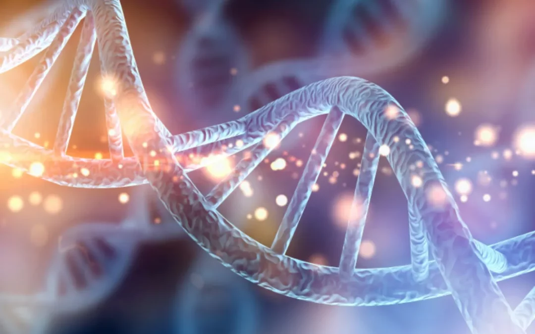
Gut-Brain Axis, Microbiome and Gut health
Your gut is more than just a digestion center—it’s the command hub for your immune system, mood, metabolism, and even skin health. When your gut is out of balance, it can affect nearly every system in your body. Recognizing the signs of an unhealthy gut is the first step toward restoring balance and improving overall well-being.
This comprehensive guide will help you identify symptoms of poor gut health, understand underlying causes, and explore natural remedies to heal your digestive system.
Why Gut Health Matters
Your gastrointestinal tract contains trillions of bacteria, fungi, and other microorganisms collectively known as the gut microbiome. A healthy gut promotes:
- Efficient digestion and nutrient absorption
- Immune regulation
- Hormone production (like serotonin and melatonin)
- Detoxification
When this complex system becomes imbalanced, it can lead to a wide range of symptoms and chronic conditions.
Common Signs of an Unhealthy Gut
The symptoms of poor gut health often extend beyond digestion. Here’s what to watch for:
1. Digestive Issues
- Bloating
- Gas or belching
- Constipation or diarrhea
- Heartburn or acid reflux
- Food intolerances or sensitivities
2. Fatigue and Brain Fog
- Low energy even after adequate sleep
- Trouble concentrating
- Forgetfulness or confusion
3. Skin Conditions
- Acne
- Eczema
- Psoriasis
- Rosacea
4. Autoimmune Conditions
- Hashimoto’s thyroiditis
- Rheumatoid arthritis
- Inflammatory bowel diseases (Crohn’s, ulcerative colitis)
5. Mood and Mental Health Problems
- Anxiety
- Depression
- Irritability or mood swings
6. Frequent Illness
- Colds, sinus infections, and flus
- Slower recovery from sickness
- Chronic low-grade inflammation
7. Unexplained Weight Changes
- Weight gain despite diet and exercise
- Difficulty losing weight
- Unstable blood sugar levels
8. Sugar Cravings and Poor Appetite Regulation
- Constant cravings for sweets or carbs
- Feeling hungry soon after eating
9. Bad Breath or Coated Tongue
- Halitosis
- White or yellow film on the tongue (may indicate candida overgrowth)
10. Nutrient Deficiencies
- Anemia (iron or B12 deficiency)
- Weak nails and hair loss
- Dry skin or poor wound healing
Causes of an Unhealthy Gut
Understanding the root causes can help you correct imbalances and support gut repair.
Major Contributors:
- Processed foods and sugar: Promote bad bacteria and inflammation
- Antibiotics and NSAIDs: Harm beneficial gut flora and lining
- Chronic stress: Disrupts gut-brain axis and microbiome
- Lack of fiber: Starves healthy bacteria
- Sleep deprivation: Weakens immune defenses and slows gut repair
- Toxins: Pesticides, BPA, heavy metals
- Infections: Candida, SIBO, parasites
How to Support a Healthy Gut
Healing your gut requires a holistic approach that includes dietary changes, stress management, and lifestyle improvements.
1. Eat a Gut-Friendly Diet
- Eliminate processed foods, sugar, and alcohol
- Eat high-fiber vegetables, fruits, nuts, and seeds
- Include fermented foods like kefir, sauerkraut, kimchi, and yogurt
- Choose anti-inflammatory foods: leafy greens, turmeric, ginger, salmon
2. Take Targeted Supplements
- Probiotics: Restore beneficial gut bacteria
- Prebiotics: Feed healthy microbes (inulin, chicory, garlic, onions)
- Digestive enzymes: Support better breakdown and absorption
- L-glutamine: Helps repair the gut lining
3. Manage Stress
- Practice deep breathing or meditation
- Spend time in nature
- Exercise regularly, but avoid overtraining
4. Improve Sleep Quality
- Aim for 7–9 hours of restful sleep
- Avoid screens before bed
- Maintain a consistent sleep-wake cycle
5. Avoid Toxins
- Choose organic foods when possible
- Filter drinking water
- Use natural personal care and cleaning products
Expert Insight: What Functional Practitioners Say
Dr. Amy Myers, author of The Autoimmune Solution, explains:
“A healthy gut is essential for optimal immune function, brain health, and weight control. Healing the gut is foundational to wellness.”
Dr. Josh Axe adds:
“If you’re experiencing chronic symptoms, it’s likely tied to your gut. Repairing the gut is one of the best investments in long-term health.”
Frequently Asked Questions (FAQs)
Can gut problems cause anxiety or depression?
Yes. The gut-brain axis plays a significant role in mood regulation. Gut dysbiosis can affect neurotransmitter production and contribute to mental health disorders.
Is bloating always a sign of an unhealthy gut?
Not always, but persistent bloating often indicates poor digestion, food intolerance, or microbiome imbalance.
How long does it take to heal an unhealthy gut?
Most people see improvements within 4–12 weeks of consistent changes, though full healing may take several months.
Should I take probiotics every day?
In many cases, yes—especially after antibiotic use. Choose a high-quality supplement with diverse strains.
Final Thoughts: Listen to your Gut
Your body often gives subtle—and not-so-subtle—clues when something’s wrong in your digestive system. If you’re experiencing several signs of an unhealthy gut, it may be time to make changes that support healing from the inside out.
With the right combination of nutrition, supplements, stress management, and sleep, you can restore your gut and experience lasting improvements in energy, immunity, mood, and skin.
Disclaimer: This article is for informational purposes only and does not replace medical advice. Consult a qualified healthcare provider for personalized guidance.
Learn the key signs of an unhealthy gut, what causes poor gut health, and how to heal your gut naturally for better overall wellness.

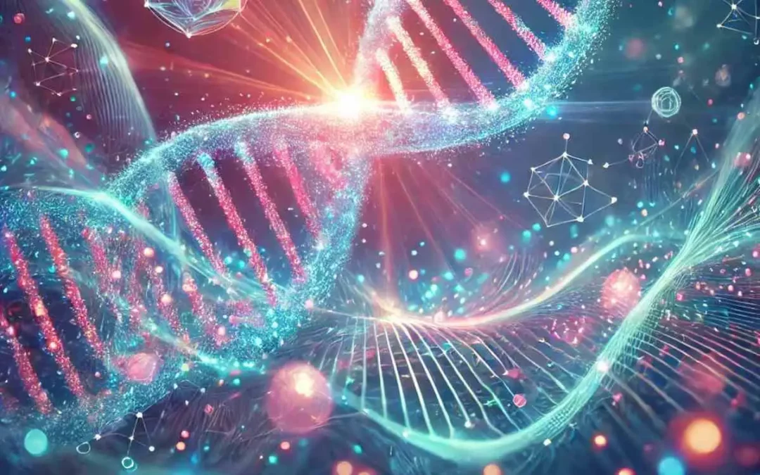
Gut-Brain Axis, Microbiome and Gut health
Leaky Gut Syndrome—also known as increased intestinal permeability—is a condition that has gained growing attention in the fields of integrative and functional medicine. Though not yet widely recognized by conventional medicine as a formal diagnosis, many health experts believe it plays a significant role in various chronic conditions, particularly those involving the immune system, digestive health, and inflammation.
In this article, we’ll explore what leaky gut syndrome is, what causes it, how it affects the body, and what you can do to heal your gut naturally.
What Is Leaky Gut Syndrome?
Your digestive tract is lined with a thin layer of epithelial cells forming a barrier known as the intestinal lining. This barrier is designed to absorb nutrients while preventing harmful substances like toxins, microbes, and undigested food particles from entering your bloodstream.
In leaky gut syndrome, this protective barrier becomes compromised. The tight junctions between intestinal cells loosen, allowing foreign particles to “leak” into the bloodstream. This can trigger inflammation and immune responses, contributing to a wide range of health issues.
Common Symptoms of Leaky Gut
Leaky gut can manifest differently from person to person, but common symptoms include:
-
Digestive issues: bloating, gas, diarrhea, constipation, IBS-like symptoms
-
Food sensitivities: new or worsening intolerances to gluten, dairy, soy, etc.
-
Brain fog and poor concentration
-
Chronic fatigue
-
Joint pain or arthritis-like symptoms
-
Headaches or migraines
-
Skin issues: eczema, acne, rosacea, psoriasis
-
Autoimmune conditions: Hashimoto’s, lupus, rheumatoid arthritis
-
Mood imbalances: anxiety, depression
Note: These symptoms can overlap with other health conditions, so it’s important to seek proper evaluation from a qualified healthcare provider.
What Causes Leaky Gut?
There isn’t a single cause of leaky gut; rather, it’s usually a combination of factors that damage the intestinal lining over time. The most common triggers include:
1. Poor Diet
-
High intake of processed foods, refined sugar, and unhealthy fats
-
Gluten and casein (proteins found in wheat and dairy) may contribute in sensitive individuals
-
Excessive alcohol consumption
2. Chronic Stress
3. Imbalance of Gut Microbiome (Dysbiosis)
4. Toxin Exposure
5. Use of Certain Medications
6. Infections and Chronic Inflammation
-
Pathogens such as H. pylori or parasites, and conditions like IBD (Crohn’s, ulcerative colitis), can damage the intestinal lining
How Is Leaky Gut Diagnosed?
While there’s no standardized test specifically for leaky gut, functional medicine practitioners may use:
How to Heal a Leaky Gut Naturally
Healing a leaky gut involves removing the irritants, restoring the gut barrier, and replenishing the gut microbiome. This approach is often summarized by the “4R Program”:
1. Remove
Eliminate foods and factors that irritate the gut:
-
Gluten, dairy, processed sugar, alcohol
-
Food allergens and sensitivities
-
Inflammatory medications (if possible)
2. Replace
Support digestion with:
3. Reinoculate
Rebuild healthy gut flora:
-
Probiotics (look for strains like Lactobacillus, Bifidobacterium)
-
Fermented foods: sauerkraut, kimchi, kefir, yogurt (if tolerated)
4. Repair
Support gut lining regeneration with:
Best Foods for Leaky Gut
Incorporate whole, anti-inflammatory foods that nourish the gut lining:
-
Bone broth
-
Wild-caught fish (rich in omega-3s)
-
Leafy greens and non-starchy vegetables
-
Berries and low-glycemic fruits
-
Coconut products
-
Sprouted seeds (chia, flax)
-
Fermented foods
-
Healthy fats (avocado, olive oil)
Avoid common gut irritants like processed foods, sugar, gluten, dairy (if intolerant), and artificial additives.
Lifestyle Changes That Support Gut Health
-
Manage stress: yoga, meditation, deep breathing
-
Get enough sleep: 7–9 hours per night
-
Exercise regularly: supports digestion and reduces inflammation
-
Stay hydrated: water is essential for healthy digestion
-
Avoid smoking and excess alcohol
Is Leaky Gut Real? The Medical Debate
While many functional and integrative practitioners support the concept of leaky gut, mainstream medicine often remains skeptical due to limited large-scale studies. However, intestinal permeability is a well-documented scientific phenomenon, especially in conditions like celiac disease, Crohn’s disease, and IBS.
Emerging research suggests that gut barrier dysfunction may play a role in:
More studies are needed, but anecdotal and clinical evidence supports the idea that healing the gut can improve a variety of chronic symptoms.
When to See a Doctor
If you’re experiencing chronic digestive problems, food sensitivities, or signs of systemic inflammation, consult with a:
Testing and personalized guidance can help determine the best course of action.
Final Thoughts
Leaky gut syndrome may be at the root of many common but poorly understood health complaints. While the medical community continues to explore its significance, there’s a growing consensus that gut health is central to overall wellness.
By adopting a gut-healing lifestyle—focused on clean nutrition, stress reduction, and targeted supplementation—you can support your digestive system and promote long-term health.
Resources & References
-
Fasano A. “Zonulin and its regulation of intestinal barrier function: the biological door to inflammation, autoimmunity, and cancer.” Physiological Reviews, 2011.
-
Methylation
Methylation is one of the most important biochemical processes in the body — yet it’s often overlooked. It influences everything from gene expression and detoxification to mood regulation and cardiovascular health. When methylation is impaired, symptoms like fatigue, anxiety, poor concentration, and even inflammation may arise. The good news? Specific nutrients can support healthy methylation and help your body function at its best.
What Is Methylation?
Methylation is a chemical process that involves adding a “methyl group” (one carbon and three hydrogen atoms) to other molecules. It helps regulate gene expression, build neurotransmitters, process hormones, and detoxify harmful substances. Methylation also plays a role in cellular energy production and immune function.
Signs of Poor Methylation
- Fatigue or brain fog
- Hormonal imbalances
- Elevated homocysteine levels
- Mood disorders (e.g., anxiety, depression)
- Detox issues or chemical sensitivity
Key Nutrients of Methylation Support
1. Methylfolate (5-MTHF)
- Active form of folate that bypasses common genetic mutations like MTHFR
- Supports DNA synthesis and neurotransmitter production
2. Methylcobalamin (Vitamin B12)
- Essential for methylation and red blood cell formation
- Supports nervous system health
3. Vitamin B6 (P5P)
- Needed to convert homocysteine into cysteine
- Supports neurotransmitter balance
4. Betaine (Trimethylglycine)
- Donates methyl groups to support homocysteine clearance
- Found in beets and whole grains
5. Riboflavin (Vitamin B2)
- Cofactor for MTHFR enzyme
- Enhances folate metabolism
6. Magnesium
- Involved in over 300 enzymatic reactions, including those related to methylation
Choosing the Right Supplement
- Look for bioavailable forms (methylfolate, methylcobalamin, P5P)
- Avoid synthetic folic acid if you have MTHFR variants
- Use targeted blends that combine key cofactors
- Start low and monitor response, especially if sensitive
Final Thoughts
Optimizing your methylation is one of the smartest steps you can take for long-term wellness. Whether you’re looking to boost mood, support detox, or protect your DNA, the right combination of nutrients can make a big difference. Work with a knowledgeable practitioner to personalize your protocol and monitor your progress.
Mitochondrial health
Mitochondria are the powerhouse of your cells — quite literally. They generate ATP, the energy currency your body needs for everything from brain function to muscle movement. But when your mitochondria aren’t working well, you may feel fatigued, foggy, and older than your years. The good news? Mitochondrial function can be supported and even improved through diet, lifestyle, and targeted supplementation. Here’s how.
Why Mitochondrial Health Matters
Healthy mitochondria are essential for:
- Energy production (ATP)
- Metabolic efficiency
- Brain and heart function
- Hormone synthesis
- Cellular repair and regeneration
Mitochondrial dysfunction is linked to aging, chronic fatigue, neurodegenerative diseases, and metabolic disorders. Supporting your mitochondria helps you feel energized, focused, and resilient.
Signs of Mitochondrial Dysfunction
- Low energy or chronic fatigue
- Brain fog or memory problems
- Muscle weakness or slow recovery
- Sensitivity to stress or poor stress tolerance
- Mood swings or depression
Natural Strategies to Boost Mitochondrial Function
1. Eat Mitochondria-Friendly Foods
- Dark leafy greens, berries, nuts, and fatty fish
- Nutrient-dense whole foods that provide antioxidants and co-factors
2. Support with Key Nutrients
- CoQ10: Essential for electron transport and ATP production
- Acetyl-L-Carnitine: Transports fatty acids into mitochondria
- Alpha-lipoic acid: Regenerates antioxidants and supports energy
- Magnesium and B vitamins: Critical for energy metabolism
3. Practice Intermittent Fasting or Time-Restricted Eating
- Supports mitochondrial biogenesis and autophagy (cellular cleanup)
4. Engage in Regular Exercise
- Particularly aerobic and resistance training stimulate mitochondrial growth and function
5. Reduce Toxin Exposure
- Minimize environmental toxins, processed foods, and oxidative stressors
6. Prioritize Quality Sleep
- Mitochondria repair during deep sleep phases; poor sleep = poor cellular health
Advanced Tools for Mitochondrial Support
- Red light therapy (photobiomodulation)
- Cold exposure or contrast therapy
- NAD+ boosting compounds (e.g., nicotinamide riboside, NMN)
Final Thoughts
Your mitochondria are at the core of your energy, mood, and longevity. Small changes in diet and lifestyle can make a big difference in how your cells produce energy and recover from stress. Support your mitochondria, and your entire body will thank you.

Detoxification, Immunity, Weight loss
Juice cleanse has surged in popularity as a quick fix for detox, weight loss, and a “reset” for the body. But what actually happens when you consume nothing but juice for several days? Is it a powerful health boost—or a risky shortcut?
In this article, we’ll break down the science behind juice cleanses, the short-term effects you might experience (both good and bad), and the long-term implications of replacing solid food with liquid nutrition.
What Is a Juice Cleanse?
A juice cleanse is a short-term dietary regimen where you consume only fruit and vegetable juices—often raw and cold-pressed—for a period ranging from 1 to 10 days. Many commercial juice cleanse programs promise to flush toxins from the body, improve digestion, boost immunity, and promote weight loss.
But what does science say? While some claims are rooted in nutritional truth, others are exaggerated or misleading.
Day-by-Day Breakdown: What Happens to Your Body?
Day 1: The Sugar Surge
Your body is used to solid food. On the first day, as you switch to juices—especially fruit-based ones—you experience a spike in blood sugar. Even natural sugars can trigger insulin fluctuations, leading to:
Day 2–3: Detox or Depletion?
This is often the hardest phase. As your glycogen stores begin to deplete, your body shifts into a mild catabolic state:
-
Fatigue sets in
-
You may feel irritable or foggy-headed
-
Some people experience nausea, dizziness, or digestive upset
-
However, some report a “light” feeling, reduced bloating, and even better sleep
Day 4–5: Adaptation and Ketosis
If your cleanse continues beyond three days, your body starts adjusting:
-
You may enter mild ketosis, burning fat for energy
-
Energy levels may stabilize (unless the calorie intake is too low)
-
Hunger often diminishes
-
Nutrient deficiencies can start to emerge, especially if the juice mix lacks diversity (e.g., no leafy greens, protein sources, or healthy fats)
The Pros of a Juice Cleanse
When done mindfully and for a limited time, a juice cleanse may offer several benefits:
✅ Increased Micronutrient Intake
Fresh juices can be nutrient-dense, particularly in vitamins A, C, and K, folate, and plant antioxidants.
✅ Hydration Boost
Juices contribute to overall hydration, especially if you’re including cucumber, celery, and watermelon-based blends.
✅ Short-Term Weight Loss
A caloric deficit is inevitable. While some of this is water weight, it can jumpstart a longer-term lifestyle change.
✅ Mental Reset
Eliminating processed foods—even temporarily—can help break habits, reduce cravings, and renew motivation.
The Cons and Risks You Should Know
Despite the hype, juice cleanses come with downsides—especially if prolonged or repeated often.
❌ Blood Sugar Spikes
Fruit-heavy juices can contain more sugar than a can of soda, spiking insulin and potentially stressing your pancreas.
❌ Lack of Protein and Fat
Protein is essential for muscle maintenance, enzyme function, and immune health. Most juice cleanses lack complete amino acids and healthy fats, leading to:
-
Muscle loss
-
Fatigue
-
Weakened immunity
❌ Gut Microbiome Disruption
Fiber plays a vital role in feeding beneficial gut bacteria. Juice removes nearly all insoluble fiber, which may disturb your gut flora balance.
❌ Metabolism Slowdown
A prolonged calorie deficit can slow down your metabolic rate, making it harder to sustain weight loss after the cleanse.
What About Detoxification?
One of the most widely touted claims of juice cleanses is “detox.” But the truth is: your body already has a highly effective detox system.
The liver, kidneys, lungs, and digestive system are constantly working to remove toxins. There’s no scientific evidence that juice cleanses “flush out” more toxins than these systems already do naturally.
However, reducing the load on your digestive system (by eliminating processed foods, alcohol, and excess sugar) can support these organs. That said, whole foods—especially fiber-rich ones—may do the job even better.
Who Should Avoid Juice Cleanse?
Juice cleanses are not recommended for:
-
Pregnant or breastfeeding women
-
People with diabetes or hypoglycemia
-
Those with kidney disease (due to high oxalate content)
-
Children and teenagers
-
Individuals with eating disorders or a history of disordered eating
Always consult a healthcare provider before starting a cleanse—especially if you have an underlying condition.
Tips for a Healthier Juice Cleanse (If You Still Want to Try It)
-
Limit it to 1–3 days max
-
Include mostly vegetables (spinach, kale, cucumber) and limit high-sugar fruits
-
Add fiber supplements or eat fiber-rich soups on the side
-
Drink plenty of water
-
Break the cleanse gradually by reintroducing light whole foods (soups, steamed veggies, oats)
The Bottom Line: Is It Worth It?
A short juice cleanse can serve as a psychological and dietary reset—but it’s not a miracle fix, nor a sustainable approach to health.
Rather than eliminating solid foods, a more balanced strategy is to incorporate more fresh juices alongside fiber-rich, whole-food meals. Focus on long-term lifestyle shifts instead of quick-fix detoxes.
Quick Summary
| Pros |
Cons |
| Increased vitamins & antioxidants |
Low in protein and fiber |
| Hydration and reduced cravings |
Blood sugar fluctuations |
| Short-term weight loss |
Muscle loss, fatigue |
| Possible mental reset |
Not suitable for everyone |
Remember: Health is a marathon, not a sprint. Drink your greens—but don’t forget to chew your food too.
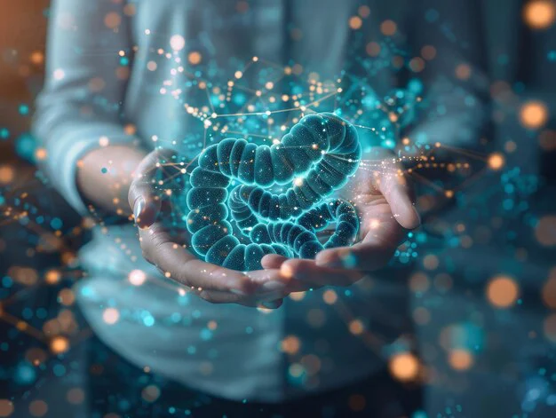
Gut-Brain Axis, Microbiome and Gut health
Gut health is becoming one of the most discussed topics in wellness—and for good reason. A well-functioning digestive system doesn’t just help with regularity and nutrient absorption; it plays a central role in your immune function, brain health, hormonal balance, and even emotional well-being. For health-conscious adults looking to improve their overall vitality, optimizing gut health is a foundational step that pays long-term dividends.
In this guide, we’ll walk you through what gut health really means, how the microbiome affects your daily life, and step-by-step strategies you can begin today to support a more balanced and vibrant digestive ecosystem.
What Is Gut Health and Why Does It Matter?
Your gut is home to a vast and dynamic ecosystem known as the gut microbiome. This collection of bacteria, fungi, viruses, and other microbes performs essential tasks that influence nearly every system in the body. A well-balanced microbiome can:
-
Break down complex carbohydrates and fibers into usable nutrients
-
Synthesize key vitamins like B12, K2, and folate
-
Protect against pathogens and harmful bacteria
-
Communicate with your brain via the gut-brain axis
-
Regulate inflammation throughout the body
When your gut microbiome is out of balance—a condition known as dysbiosis—you may experience:
-
Bloating, gas, or irregular bowel movements
-
Food intolerances or sensitivities
-
Skin issues like eczema or acne
-
Brain fog, fatigue, and even low mood or anxiety
-
A weakened immune response and more frequent illness
Luckily, research shows that with targeted dietary and lifestyle changes, you can significantly improve your gut health in just a few weeks.
How to Improve Gut Health: Step-by-Step
1. Prioritize a High-Fiber Diet (Prebiotic-Rich Foods)
Fiber is the fuel for your friendly gut bacteria. When these microbes ferment fiber, they produce short-chain fatty acids (SCFAs)—powerful anti-inflammatory compounds that heal the gut lining and support immunity.
Include at least 25–35g of fiber per day from sources like:
-
Vegetables: broccoli, carrots, leafy greens, leeks
-
Fruits: apples, berries, bananas (slightly green for resistant starch)
-
Whole grains: oats, buckwheat, barley, bulgur
-
Legumes: lentils, black beans, chickpeas
Bonus tip: Try overnight oats with chia seeds, flaxseed, and berries for a fiber-rich breakfast that feeds your microbiome.
2. Add Fermented Foods (Live Probiotics)
Fermented foods are natural sources of probiotics, or live beneficial bacteria that can replenish and diversify your gut microbiome.
Best options include:
-
Yogurt and kefir with live active cultures
-
Sauerkraut, kimchi, and miso for savory dishes
-
Kombucha as a refreshing drink
-
Tempeh and natto (great plant-based protein + probiotics)
Pro tip: Add a tablespoon of sauerkraut to your salad or sandwich daily—simple and gut-friendly!
3. Stay Well-Hydrated
Water is essential for digestion—it helps break down food, dissolve nutrients, and soften stool for easier elimination. Aim for 2 to 2.5 liters of water daily (8–10 cups), or more if you’re active.
Gut hydration booster: Try warm water with lemon in the morning to stimulate digestion and liver detox pathways.
4. Reduce Stress (Gut-Brain Axis in Action)
Stress alters gut motility, reduces enzyme production, and even changes the composition of your microbiome. Chronic stress can lead to issues like leaky gut, IBS, or food intolerances.
To calm your gut, consider:
-
Daily mindfulness meditation (5–10 min)
-
Breathing exercises (4-7-8 method)
-
Gentle movement like walking, yoga, or Tai Chi
-
Spending time in nature
Science says: A recent study showed that mindfulness-based stress reduction (MBSR) significantly improved IBS symptoms in 8 weeks.
5. Improve Sleep Hygiene
Your gut microbiome follows a circadian rhythm, just like you. Poor sleep disrupts microbial balance and weakens your immune system. Aim for:
-
7–9 hours of sleep nightly
-
Consistent sleep/wake times (even on weekends)
-
Avoid screens 1 hour before bed
-
Create a cool, dark, tech-free sleep environment
Bonus: Magnesium glycinate or herbal teas (e.g. chamomile, lemon balm) can support deeper, more restful sleep.
6. Avoid Gut Disruptors
Some common foods and habits can damage the delicate gut lining or promote overgrowth of harmful microbes. Try to limit or eliminate:
-
Ultra-processed foods and refined sugars
-
Alcohol and caffeine in excess
-
Artificial sweeteners (especially sucralose, aspartame)
-
Overuse of NSAIDs (e.g. ibuprofen), antibiotics, and proton pump inhibitors
Gut tip: If you must take antibiotics, follow up with a quality multi-strain probiotic for at least 4–6 weeks to restore balance.
7. Feed Your Gut with Prebiotics & Polyphenols
Prebiotics = food for your probiotics. Polyphenols = plant compounds that support microbial diversity and reduce gut inflammation.
Include daily:
-
Prebiotics: garlic, onions, asparagus, leeks, Jerusalem artichokes
-
Polyphenol-rich foods: green tea, dark chocolate (70%+), berries, olives, turmeric
Fun fact: The Mediterranean diet is rich in prebiotics and polyphenols, making it one of the most gut-friendly diets on earth.
Optional: Tips & Pitfalls to Avoid
✔️ Do:
-
Rotate your fiber sources weekly
-
Start slowly with fermented foods (too much = bloating)
-
Cook cruciferous veggies if raw causes gas
❌ Don’t:
-
Cut out entire food groups without guidance
-
Assume all bloating means food intolerance
-
Take random probiotics without knowing the strain
Final Thoughts
A healthy gut is not a luxury—it’s the foundation of your physical, mental, and emotional vitality. By applying the strategies above consistently, you’ll not only reduce digestive discomfort but also enhance your energy, mood, immunity, and skin health.
The best part? These changes don’t require medication—just smart food choices, mindful habits, and a little patience.

Autism and Genes, Neuroplasticity
Lithium is often associated with psychiatric medication, but at low doses, a form called lithium orotate is gaining attention for its potential neuroprotective effects. Naturally occurring in trace amounts in water and some foods, lithium plays a role in brain health, mood regulation, and cellular resilience. This article explores how lithium orotate may support cognitive function, emotional balance, and long-term brain health — and how to use it wisely.
What Is Lithium Orotate?
Lithium orotate is a salt of lithium and orotic acid, available as a nutritional supplement. Unlike prescription lithium carbonate, which is used to treat bipolar disorder at high doses, lithium orotate provides lithium in much smaller amounts — typically 1 to 5 mg per day — which may still exert therapeutic benefits with a better safety profile.
Lithium Orotate and Brain: Benefits of Low-Dose Lithium Orotate
1. Neuroprotection
- Enhances BDNF (brain-derived neurotrophic factor), supporting neuron growth and repair
- Protects against excitotoxicity and oxidative stress
2. Mood Stabilization
- May reduce anxiety, irritability, and emotional reactivity
- Supports balanced neurotransmitter activity
3. Cognitive Support
- Improves focus and working memory in some individuals
- May slow age-related cognitive decline
4. Anti-Aging and Longevity Potential
- Animal studies suggest lithium may increase lifespan and reduce age-related inflammation
Mechanisms of Action
Lithium influences multiple cellular pathways, including:
- GSK-3 inhibition: Affects mood, circadian rhythms, and neuroplasticity
- NMDA receptor modulation: Helps balance glutamate activity
- Telomere maintenance: May support cellular aging resistance
Is It Safe?
In low doses, lithium orotate is generally well tolerated. However:
- Long-term use should be monitored, especially in those with thyroid or kidney conditions
- It’s best to consult a healthcare provider before starting, particularly if taking other medications
Suggested Use and Dosage
- Typical dose: 1–5 mg elemental lithium daily (check label carefully)
- Best taken with food
- Start low and observe effects gradually
Final Thoughts
Lithium orotate is a promising tool in the natural mental wellness toolkit. From neuroprotection to mood balance, its broad mechanisms of action make it a fascinating subject of research and practice. While not a substitute for clinical treatment when needed, it may offer gentle yet meaningful support for cognitive and emotional health.
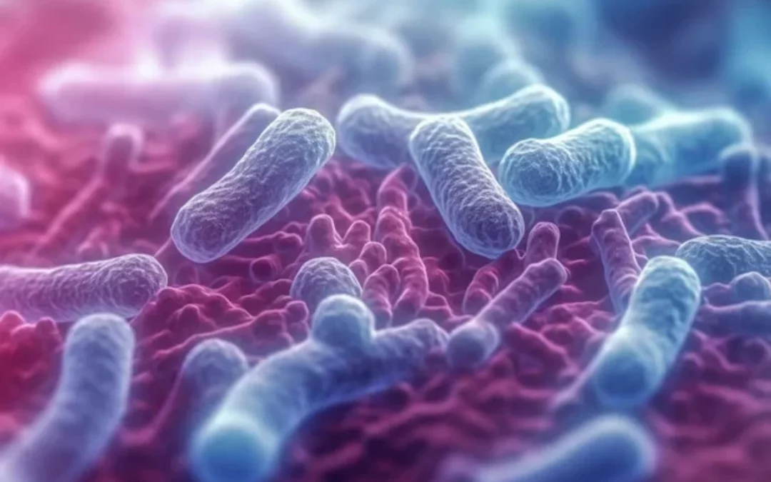
Autism and Genes, Microbiome and Gut health
Is there a link between microbiom and autism? The gut and the brain are more connected than we once thought — and this connection is especially relevant in autism spectrum disorder (ASD). Emerging research highlights how the gut microbiome may influence neurodevelopment, immune regulation, and behavior in individuals with autism. In this article, we explore the complex relationship between gut health and autism and discuss natural interventions that may support improved outcomes.
What Is the Gut Microbiome?
The gut microbiome refers to the trillions of microorganisms residing in the digestive tract. These microbes help regulate digestion, produce neurotransmitters, modulate the immune system, and maintain the integrity of the gut barrier. Dysbiosis — an imbalance in the microbiome — is increasingly associated with neurological and developmental disorders.
Microbiome Differences in Individuals with Autism
Several studies have found distinct differences in the gut microbiome composition of individuals with ASD, including:
- Lower diversity of gut bacteria
- Increased levels of Clostridium and Desulfovibrio
- Reduced beneficial strains like Bifidobacterium and Lactobacillus
These microbial shifts are linked to symptoms such as:
- Gastrointestinal distress (constipation, diarrhea, bloating)
- Irritability, anxiety, and sleep issues
- Increased immune activation and inflammation
How the Gut Microbiome Affects the Brain
The gut and brain communicate through the vagus nerve, immune signaling, and microbial metabolites (like short-chain fatty acids). Disruptions in this gut-brain axis may contribute to neuroinflammation, altered neurotransmitter balance, and behavioral changes associated with autism.
Key microbial influences include:
- Short-chain fatty acids (SCFAs): May influence brain development but can be harmful in excess
- Tryptophan metabolism: Affects serotonin production
- Lipopolysaccharides (LPS): Inflammatory bacterial byproducts that can cross the blood-brain barrier
Natural Strategies to Support Gut-Brain Health in ASD
1. Dietary Interventions
- Implement a gluten-free, casein-free (GFCF) diet if beneficial
- Avoid ultra-processed foods and additives
- Include fiber-rich, whole foods to support microbial diversity
2. Probiotic and Prebiotic Supplementation
- Probiotics such as Lactobacillus plantarum, Bifidobacterium infantis, and Saccharomyces boulardii may support gut balance
- Prebiotics like inulin and FOS can feed beneficial bacteria
3. Address Gut Inflammation and Leaky Gut
- Use nutrients like L-glutamine, zinc carnosine, and omega-3s to support gut lining integrity
4. Consider Targeted Microbiome Testing
- Stool tests can reveal specific imbalances and guide personalized protocols
Final Thoughts
The gut microbiome plays a pivotal role in neurological and behavioral health, particularly in autism spectrum disorder. While no single intervention is a cure, improving gut health through diet, supplementation, and testing can be a powerful part of a holistic support plan. As always, consult with your child’s healthcare provider before making significant changes.
Call to Action: Want to learn more about supporting your child’s gut-brain health? Download our free GFCF recipe guide or book a personalized consultation at OrganiClinic.com.