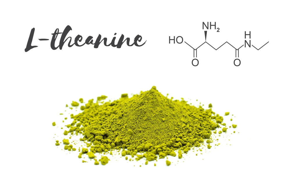
L-Theanine: Unlocking the Power of Balance in Your Brain
")
In the fast-paced world, we live in, it’s no surprise that many of us seek ways to enhance our mental health and cognitive function. Enter L-theanine, an amino acid found in tea leaves, is known for its potential to promote relaxation, sharpen focus, and improve overall brain health. But what makes L-theanine so intriguing is its unique ability to rebalance two critical neurotransmitters in the brain: GABA and glutamate.
Understanding the Neurotransmitter Duo: GABA and Glutamate
Before we delve into the wonders of L-theanine, let’s grasp the roles of GABA and glutamate in the brain. These two neurotransmitters are yin and yang, playing opposing roles to maintain equilibrium.
1. GABA (Gamma-Aminobutyric Acid): GABA is the brain’s primary inhibitory neurotransmitter. It acts like a calming agent, slowing down neural activity and promoting relaxation. When GABA levels are optimal, we feel at ease, stress is reduced, and anxiety is managed effectively.
2. Glutamate: In contrast, glutamate is the brain’s principal excitatory neurotransmitter. It revs up neural activity, aiding in concentration, learning, and memory. When glutamate is well-regulated, we experience heightened mental clarity and focus.
The Delicate Balance: GABA and Glutamate in Harmony
When GABA and glutamate are in balance, our brain functions optimally, and our mental health flourishes. However, various factors, such as stress, poor diet, and certain medical conditions, can disrupt this equilibrium. Such imbalances have been linked to mental health issues, including anxiety, depression, and even neurodevelopmental disorders like ADHD and schizophrenia.
L-Theanine to the Rescue: Rebalancing GABA and Glutamate
Studies have shed light on L-theanine’s remarkable ability to restore harmony between GABA and glutamate, offering a plethora of brain health benefits:
1. Increased GABA Levels: Research has shown that L-theanine can elevate GABA levels in the brain. A study published in the Journal of Physiological Anthropology demonstrated that L-theanine intake increased GABA activity, resulting in relaxation and a reduced stress response.
2. Inhibition of Glutamate Uptake: L-theanine can also inhibit the uptake of glutamate by the brain. By doing so, it curtails excessive glutamate levels and prevents overexcitation of neural pathways. A study published in the Journal of Food Science supported this finding, indicating that L-theanine’s glutamate-blocking effect contributes to a calmer mental state.
3. Enhancement of Alpha Brain Waves: Alpha brain waves are associated with a state of relaxation, mental clarity, and focus. L-theanine has been found to enhance alpha wave activity in the brain, fostering a sense of calm alertness. A study in the journal Nutrients highlighted this effect, suggesting that L-theanine could aid in stress reduction and cognitive performance.
Studies Supporting L-Theanine's Brain Benefits
1. L-Theanine reduces psychological and physiological stress responses
https://www.ncbi.nlm.nih.gov/pmc/articles/PMC4728665/
2. L-Theanine increases GABA in the brain: the first screening in vivo
https://pubmed.ncbi.nlm.nih.gov/18296328/
3. Effects of L-Theanine Administration on Stress-Related Symptoms and Cognitive Functions in Healthy Adults: A Randomized Controlled Trial
https://pubmed.ncbi.nlm.nih.gov/31936666/
4. L-theanine, a natural constituent in tea, and its effect on mental state
Unlocking the Full Potential of L-Theanine
While L-theanine shows tremendous promise in rebalancing GABA and glutamate, it’s essential to remember that individual responses may vary. As with any supplement, consult with a healthcare professional before incorporating L-theanine into your routine, especially if you have pre-existing medical conditions or are taking medications.
Incorporating mindful practices, a balanced diet, and regular exercise alongside L-theanine may offer holistic support for your brain health. Whether you’re looking to manage stress, enhance focus, or simply achieve mental clarity, L-theanine might just be the key to unlocking the power of balance in your brain. So, go ahead, sip that soothing cup of tea, and embrace the tranquility L-theanine brings.
Resources


 (1)")

 (1)")